
Our Mission
In the Fitzpatrick Lab, we leverage the latest developments in cryo-electron microscopy with complementary biophysical techniques (proteomics, light-microscopy, microfluidics) to explore the molecular and structural basis of neurodegeneration and memory. To this end, we determine the structure and behavior of amyloid fibrils, purified directly from postmortem human brain tissue, that are implicated in a range of neurodegenerative diseases and explore their interactome.
Additionally, we aim to understand the role of protein aggregation in vivo by visualizing the cellular changes that occur in response to the formation, clearance and spread of fibrillar inclusions using light-microscopy and cryo-electron tomography. We are in the Department of Biochemistry and Molecular Biophysics and the lab is currently located on Columbia’s Manhattanville Campus, in the Jerome L. Greene Science Center.
Research Highlights

Homotypic fibrillization of TMEM106B
Cryo-EM and mass spectrometry-based proteomics of insoluble amyloid fibrils derived from postmortem human brains afflicted with diverse neurodegenerative diseases reveals widespread fibrillization of an endolysosomal membrane protein, TMEM106B, pointing toward a potentially pathogenic commonality between distinct proteinopathies.
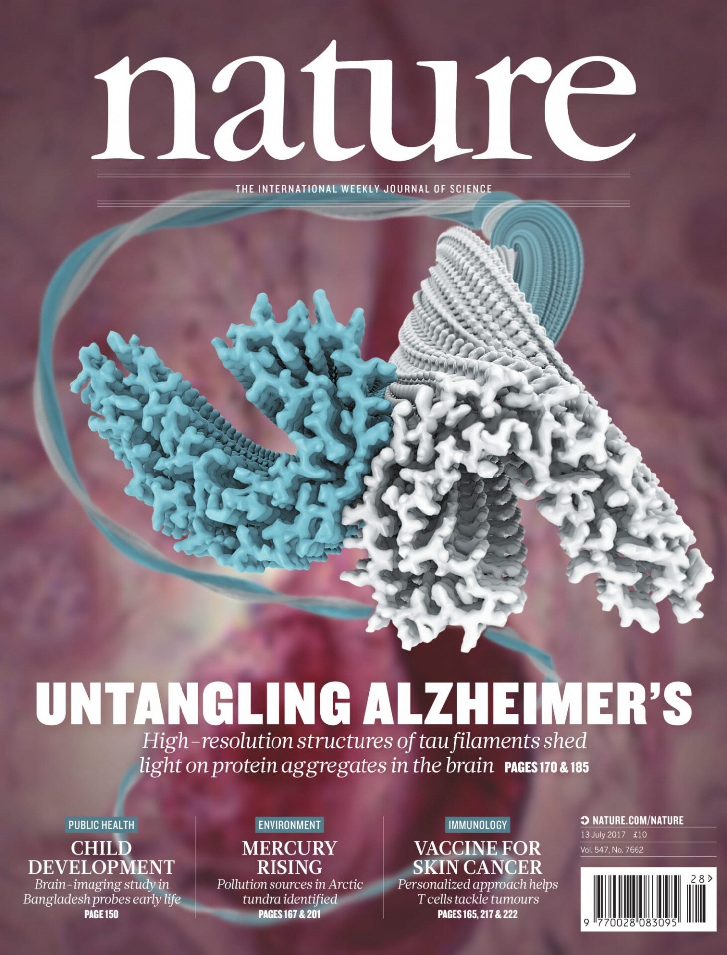
Revealing Alzheimer’s
At Columbia’s Zuckerman Institute, Anthony Fitzpatrick, PhD, traces the molecular building blocks of Alzheimer’s, Parkinson's and other devastating diseases using Nobel prize-winning cryo-electron microscopy (cryo-EM).
Structures of Human Brain-Derived Amyloid Fibrils
Many debilitating human disorders, including Alzheimer’s and Parkinson’s diseases, are associated with the conversion of normally soluble functional proteins into highly ordered aggregates known as amyloid fibrils. Determining the atomic structures of these filamentous aggregates is crucial to understanding their formation, clearance and spreading in the human body. The Fitzpatrick Lab uses cryo-EM to solve the structures of filaments isolated from postmortem brain tissue of patients – Patient-Based Structural Biology – with a range of neurological disorders to elucidate the molecular and structural basis of neurodegeneration, enabling structure-based drug design and the development of biomarkers for early detection.
News Highlights

Fitzpatrick Lab awarded CZI Visual Proteomics Grant
The Chan Zuckerberg Initiative (CZI) announced nearly $28 million in grants to support technology developments that will allow researchers to see the inner workings of cells at near-atomic resolution through next-generation electron microscopy. The Frontiers of Imaging initiative, part of CZI’s broader Imaging program, supports the development of disruptive imaging technologies that connect biological scales across organs, cells, and proteins, allowing researchers to directly visualize biological processes at the necessary resolution and context to obtain a mechanistic understanding of health and disease.
The Fitzpatrick lab will develop a pulsed ponderomotive phase plate that allows in-focus phase contrast of cryo-EM specimens. By focusing a pulsed laser to intersect the post-specimen electron beam, the ponderomotive potential of the focused laser crossover produces a scattering-angle-dependent phase shift in the electrons, resulting in a highly tunable contrast transfer function, with significantly enhanced contrast of the cells under inspection. This powerful approach to boosting contrast in cellular cryo-electron tomograms has the potential to revolutionize our understanding of biology by mapping cell atlases in unprecedented detail.

Fitzpatrick Lab undergraduates publish in Cell
The Fitzpatrick Lab is immensely proud to share that our two undergraduate research fellows, Tamta Arakhamia and Christina E. Lee, have published as co-first authors in Cell!
The study, published today in Cell, employed two complementary techniques to map the structure of tau and decipher the effects of additional molecules, called post-translational modifications (PTMs), on its surface. These new structural insights could accelerate the fight against neurodegenerative diseases, by helping researchers identify new biomarkers that detect these disorders before symptoms arise and design new drugs that target specific PTMs, preventing the onset of disease before it wreaks havoc on the brain.
Dr. Anthony Fitzpatrick and Collaborators Awarded $8.8M to Identify New Targets Against Brain Disease
Using cryo-electron microscopy (cryo-EM), Dr. Fitzpatrick’s team will focus their efforts primarily on tau tangles, clumps of misfolded tau protein that have been implicated in a host of diseases, most notably Alzheimer’s disease and chronic traumatic encephalopathy, or CTE. By reconstructing the structure of tau tangles in these diseases — atom by atom — the researchers hope to gain new insight into how tangles form, how they grow and how they drive disease progression. These findings will enable researchers to design drug molecules that prevent tangle buildup. They will also aid development of new biomarkers that detect the disease early, before symptoms arise.
Dr. Fitzpatrick will serve as lead principal investigator of this five-year project, which will be administered by the National Institute of Neurological Disorders and Stroke (NINDS). He will be joined by Leonard Petrucelli, PhD, Chair and Professor of Neuroscience at Mayo Clinic’s Florida campus, and Judith Steen, PhD, Associate Professor of Neurobiology at Harvard University and Boston Children’s Hospital.